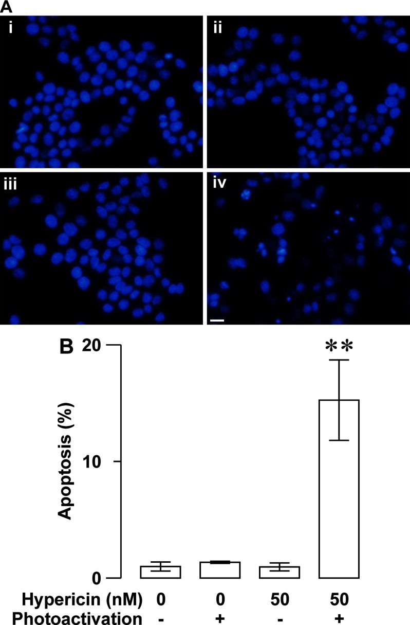
Figure 6. Photoactivated hypericin-induced up-regulation of RINm5F insulinoma cell apoptosis revealed by DAPI-staining analysis.
(A) Sample DAPI fluorescence images were acquired from specimens subjected to solvent control treatment (control), hypericin loading (hypericin), solvent control treatment followed by photoactivation (control/photoactivation) and hypericin loading followed by photoactivation (hypericin/photoactivation) respectively. A hypericin/photoactivation-treated specimen (iv) showed several apoptotic profiles characterized by chromatin condensation, nuclear shrinkage and apoptotic body formation, but control- (i), hypericin- (ii), control/photoactivation-treated specimens (iii) rarely displayed apoptotic cells. (B) Quantifications of the percentage of apoptotic cells in control, hypericin, control/photoactivation and hypericin/photoactivation groups. The percentage of apoptotic cells is very low in control (N=3), hypericin (N=3) and control/photoactivation (N=3) groups, but significantly increased in hypericin/photoactivation group (N=3). **P<0.01 compared with control, control/photoactivation and hypericn. Bar=20 μm.

