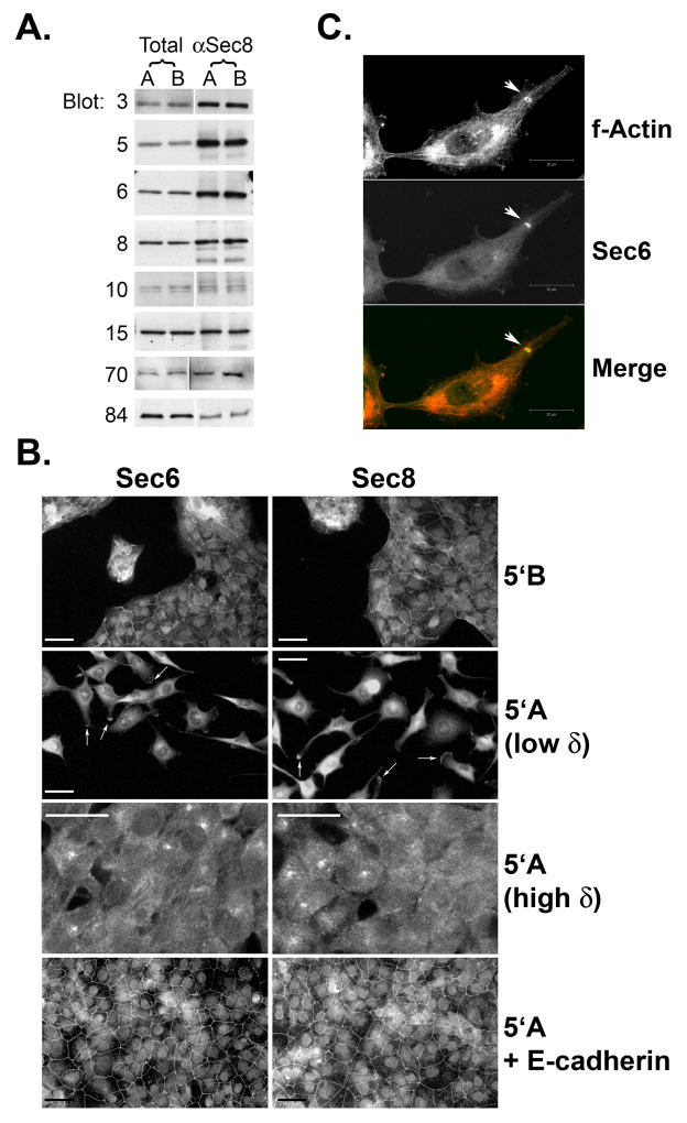
Figure 1. Exocyst expression, assembly and localization in non-metastatic and metastatic prostate tumor cells.
(A) Dunning rat R3327-5′A (“A”) and R3327–5′B (“B”) cells were extracted in 1% Triton X-100. Extracts were subjected to immunoprecipitation with antibodies to Sec8. Presence of Sec3, Sec5, Sec6, Sec8, Sec10, Sec15, Exo70 and Exo84 in equivalent amounts of whole cell extracts (“total”) and precipitated immune complexes (“αSec8”) was assessed by SDS-PAGE followed by immunoblotting with specific antibodies. (B) Sub-confluent cultures of R3327–5′B, R3327-5′A or R3327-5′A cells stably expressing human E-cadherin were cultured on type I collagen-coated coverslips, and then processed for immunofluorescent staining with antibodies to Sec6 or Sec8, as described in Materials and Methods. Samples were viewed with a Nikon Microphot-FX microscope (63X objective) and epifluorescent digital images were obtained using a Kodak DCS 760 digital camera. Arrows point to accumulations of Exocyst proteins in protrusive extensions of R3327-5′A cells. (C) Sub-confluent cultures of R3327-5′A cells were cultured on Matrigel-coated coverslips, and then processed for immunofluorescent staining with phalloidin (to label f-actin) and antibodies to Sec6. Arrowhead points to an accumulation of Exocyst within an actin-rich invadopodium. Scale bar = 20 μm.

