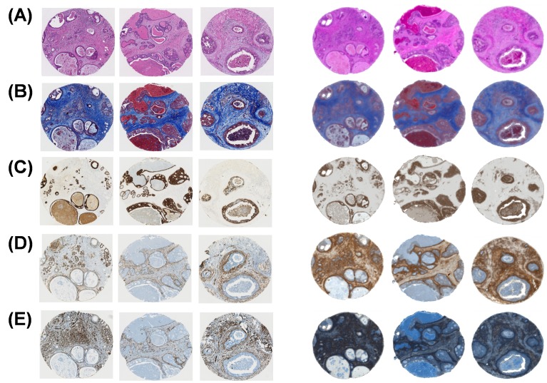
Figure 2.
Molecular imaging (three sample panel on the left) can be reproduced by chemical imaging (right panel). In addition to H&E stained images, (A) we extend the concept of stainless staining to molecularly specific stains. (B) Masson’s trichrome stain (collagen and keratin fibers). (C) High molecular weight (HMW) cytokeratin (epithelial type cell). (D) Smooth muscle alpha actin (myo-like cell). (E) Vimentin (fibroblast like cell). Each spot is 1.4 mm in diameter. Adapted with permission from World Scientific Publishing Co./Imperial College Press (Mayerich D, Walsh MJ, Kadjacsy-Balla A, Ray PS, Hewitt SM, Bhargava R. Stain-less staining for computed histopathology. Technology. 2015;3(1):27-31.)

