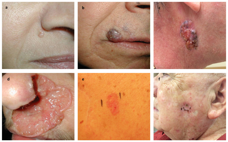
Figure 2.
Clinical variants of basal cell carcinoma. a) Nodular basal cell carcinoma with characteristic pearly surface and telangiectasias located lateral to the right alar crease. b) Pigmented variant located on the skin above the right upper lip and extending past the vermilion border into the lip. c) Large nodular basal cell carcinoma with characteristic telangiectasias and pigmented areas on the right side of the neck. d) Ulcerated aggressive basal cell carcinoma, otherwise known as “rodent ulcer.” This basal cell carcinoma spread locally into the nose, causing extensive destruction of the left nasal ala. e) Superficial basal cell carcinoma appearing as a red patch on the trunk. f) Recurrent basal cell carcinoma at the site of ED&C. (Courtesy of Samuel E. Book, MD)

