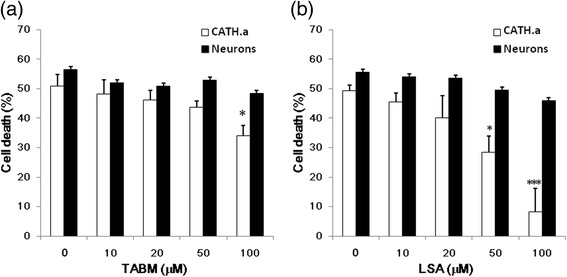
Fig. 2.

The effect of TABM and LSA on the Neurotoxicity of MPP+. CATH.a cells (opened bars) and cortical neurons (closed bars) were pretreated with various concentrations of TABM (a) or LSA (b) for 30 min, and then the cells were cultured with 250 μM MPP+ for 24 hours. Cell viability was mesured using MTT reduction assay. Results are means ± S.D. from three independent experiments. Significant differences between the cells treated with MPP+ combined with TABM or LSA were indicated by *, P < 0.05; ***, P < 0.001
