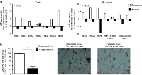
Figure 5.

Relapsed tumors show decreased expression of CD8 T cell and monocyte markers, and decreased frequencies and activity of tumor-specific CD8 T cells. Regressed, relapsed and progressed tumors as defined in Figure 3a were collected and analyzed for immune cell infiltration. (a) RNA was isolated, biotin-labeled and used to hybridize onto Illumina MouseWG-6 v2 beads. See Supplementary Text for details on sample processing and data analysis. Analyses presented in this figure were restricted to genes that correspond to T cells and monocytes. Expression of genes (annotation according to www.genecards.org) in either regressed or relapsed tumors (white and black bars, respectively) is presented as fold change in relation to progressed tumors. Calculations were based on mean values, n = 3, per tumor type. Statistical significances for both cell types were calculated with Sign tests. (b) Tumors were prepared into single cell suspensions, cultured for 4 days under T cell conditions and assessed for the presence as well as antitumor activity of TILs (n = 4). TILs were analyzed by flow cytometry for the fraction of pMHC-binding CD8 T cells within CD3 cells and by microscopic evaluation for their ability to eliminate B16 tumor cells (magnification 200×). Statistical significances were calculated with Student's t-test: *P < 0.01.
