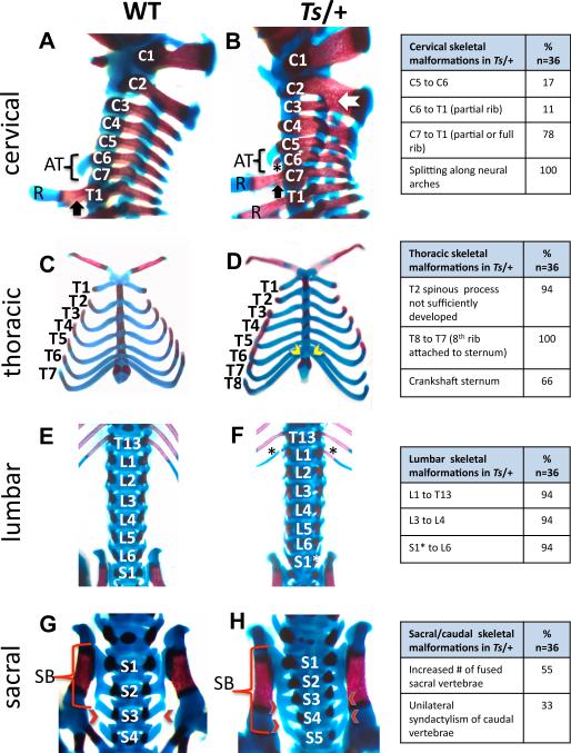
Figure 2. Homeotic transformations and skeletal patterning defects in Ts/+ embryos.
(A-H) Alizarin red and Alcian blue staining of WT and Ts/+ skeletons at E18.5. The frequency of patterning defects is presented in the table on the right (note that not all of these phenotypes are shown). Lateral view of the cervical (C) and upper thoracic (T) region. In Ts/+ embryos the anterior tuberculum (AT) normally a feature of C6 is instead present on C5. The star indicates an ectopic, partially formed rib on C6 in Ts/+ embryos. The black arrow indicates the presence of ribs (R) on the first thoracic vertebrae in Wt embryos that is a feature of C7 in Ts/+ embryos. Fusions between cervical vertebrae in Ts/+ embryos (white arrow). (C-D) Ventral view of the thorax reveals that in Ts/+ embryos there are eight pairs of vertebrosternal ribs instead of seven attached to the sternum (yellow arrow heads). The attachment site of ribs to the sternum is asymmetric in Ts/+ embryos resulting in a crankshaft sternum. (E-F) In the lumbar region, an extra pair of ribs (black asterisks) is present on the first lumbar vertebrae in Ts/+ embryos. A normal number of lumbar segments in Ts/+ embryos suggest that a partial transformation of S1 into a lumbar identity (S1*) has occurred. (G-H) Sacral vertebrae in Wt (G) and Ts/+ (H). In Wt embryos, the first three sacral vertebrae are fused to make the sacral bone (SB), while in Ts/+ embryos, the fourth and sometimes fifth sacral vertebrae are fused (red arrow heads). See also Figure S2.

