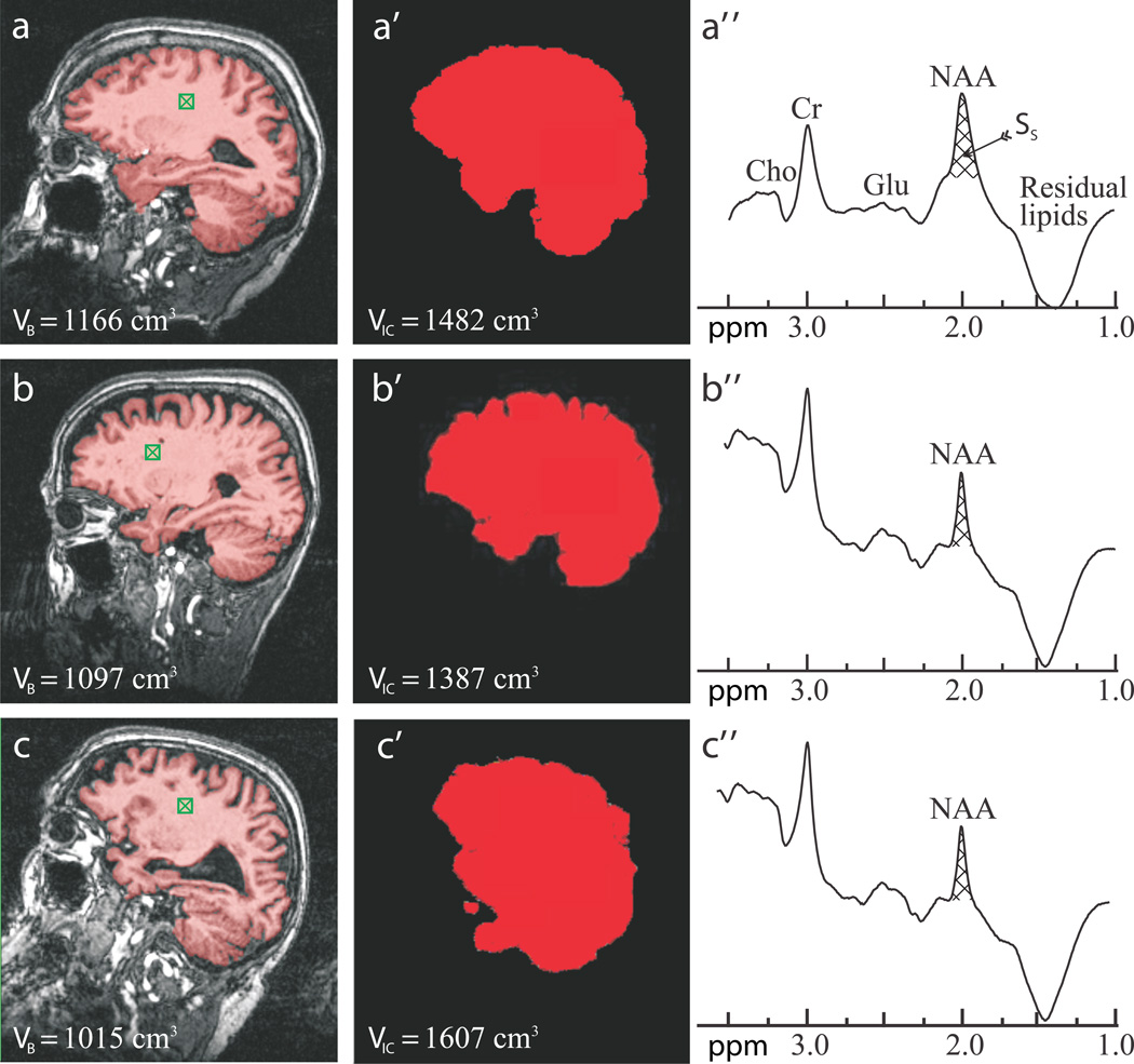
Fig. 1.
Sample FireVoxel and MRIcro segmentation used for VB and VIC (indicated on the images) and whole-head NAA spectra (not normalized for VB) from one subject from each cohort: 73 year old normal woman (a, a’, a”), 73 year old female MCI (b, b’, b”); and 80 year old male AD patient (c, c’, c”). Note the “seed” in the white matter (green hatched box in a – c), brain-capture performance of FireVoxel (a – c); intracranial volume capture of MRIcro (a’ – c’) and well defined whole-head 1H spectra (a” – c”) for straightforward integration of Ss for Eq. [1]. Note the NAA peak at 2 ppm, lipids suppression performance and that of all the other peaks in the spectrum, e.g., glutamate (Glu), total creatine (Cr) and total choline (Cho) only NAA is implicitly localized by its biochemistry to just the brain.

