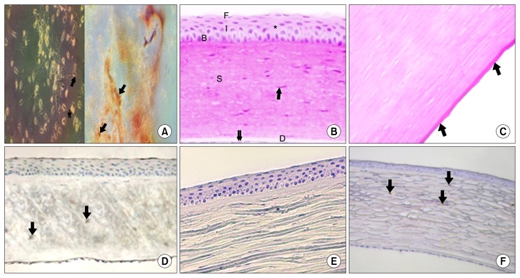
Fig. 1.
(A) BM-MSCs, on day five of isolation and culture, showing the appearance of a colony of star-shaped cells with long processes (↑), central vesicular nucleus and granular cytoplasm. Notice, a cell appears in the anaphase stage of division (*). Characterization of MSCs using CD44+ was evident as well. (B) Showing the usual layers of the cornea. The non-keratinized stratified squamous corneal epithelium (*) appears with basal columnar cells (B), intermediate polygonal cells (I) and flat surface cells (F). Thick stroma (S) containing regularly arranged fibers with sparsely distributed keratocytes (↑), homogenous Descemet’s membrane is seen (D). Notice, the simple squamous corneal endothelium (↓↓). (C) Showing PAS positive regular Descemet’s membrane (↑). (D) Showing weak alkaline phosphatase positive reaction in few corneal stromal cells (↓). (E) Showing negative immune-reaction for CD44 in the cytoplasm of epithelial and stromal cells. (F) Showing weak positive immune-reaction for vimentin in few substantia propria cells (↓). A=phase contrast inverted microscopy×200. Control group; B=H&E×250, C=PAS×560, D=Alkaline phosphatase reaction×250, E=Avidin–biotin peroxidase for CD44×250, F=Avidin–biotin peroxidase for vimentin×250.

