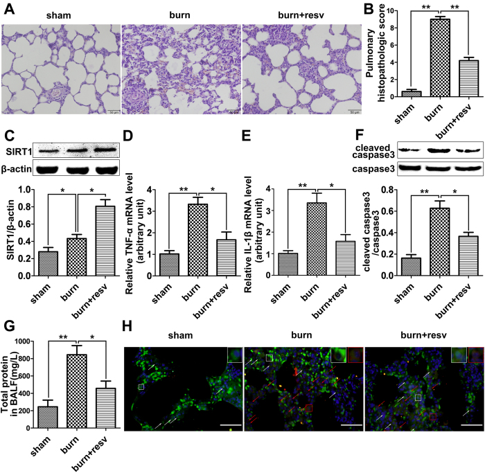
Figure 2. In vivo studies to assess the effects of resveratrol in rat lung with burn-induced ALI.
(A) H&E staining showing the morphological changes of the lung tissue from rats receiving severe burn injury at 24 h post-burn. The burn group (burn) showed increasing lung edema, alveolar hemorrhage, neutrophil infiltration, and destroyed epithelial/endothelial cell structure (middle panel) compared to the sham burn group (sham, left panel), while the resveratrol-treated burn group (burn + resv) showed significant improvement on lung morphology (right panel). Scale bar = 50 μm. (B) The pulmonary histologic score was counted at 24 h post-burn, and resveratrol significantly attenuated burn-induced high histologic score. (C) The protein level of SIRT1 was further significantly elevated by its activator resveratrol in burned rat lung. (D) Real-time PCR analysis showing the effect of resveratrol on the mRNA level of TNF-α in burned rat lung at 12 h post-treatment. (E) Real-time PCR analysis showing the effect of resveratrol on the mRNA level of IL-1β in burned rat lung at 12 h post-treatment. (F) Representative immunoblots showing the effect of resveratrol on the cleaved caspase-3 level in burned rat lung at 12 h post-treatment. (G) Total protein contents in BALF were measured in each of the three groups, and resveratrol significantly reversed the increased protein content induced by severe burn at 12 h post-treatment. (H) PMVECs positively stained with both CD31 (green) and cleaved caspase-3 (red) represent the apoptotic cells (Red arrows direct to apoptotic ECs while white arrows direct to normal ECs. Scale bar = 50 μm). Results represent mean ± SEM of six independent experiments using different rats. *p < 0.05; **p < 0.01, compared to the value in the burn group.

