Figure 4.
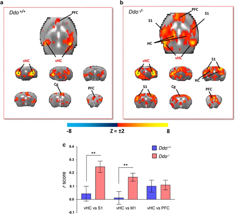
Cortico–hippocampal connectivity in Ddo−/− mice. Transverse and coronal brain section heat maps showing voxels for which the fMRI BOLD signal was significantly correlated with the ventral hippocampus (seed regions in black) in (a) Ddo+/+and (b) Ddo−/− mice. The z-score indicates the strength of the correlation. (c) A stronger and more widespread profile of cortical (somatosensory) hippocampal connectivity was apparent in Ddo−/− mice. Strength (r-score) of cortico–hippocampal connectivity in representative regions of interest of Ddo+/+ and Ddo−/− mice. **P<0.01, Student's t-test. Cg, cingulate cortex; DDO, D-aspartate oxidase; fMRI, functional magnetic resonance imaging; PFC, prefrontal cortex; S1, somatosensory (parietal) cortex; vHc, ventral hippocampus; V1, visual cortex.

