Figure 5.
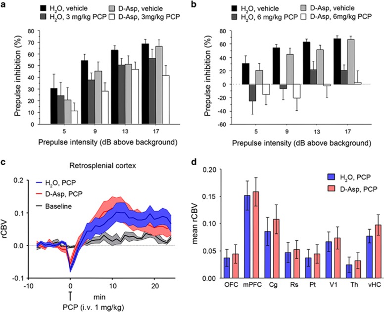
Effect of D-aspartate (D-Asp) supplementation on PCP-induced responses. (a and b) Prepulse inhibition responses to PCP after 1-month oral administration of D-aspartate in mice treated with (a) 3 mg kg−1 PCP (n=10 H2O; n=9 D-Asp) or vehicle (n=10 H2O; n=8 D-Asp), (b) 6 mg kg−1 PCP (n=10 H2O; n=10 D-Asp) or vehicle (n=10 H2O; n=8 D-Asp). Percentage of the PPI was used as dependent variable and measured at different prepulse intensities (shown as dB above 65 dB background level). All the values are expressed as the mean±s.e.m. (c and d) PCP-induced fMRI response in D-Asp- and H2O-treated adult C57BL6/J mice. In both groups of animals, PCP elicited robust and sustained cortico–limbo–thalamic fMRI activation (left and right panels). No statistically significant difference in the inter-group response to PCP was observed either at the voxel level (z>1.6, cluster corrected at P<0.01) or when integrated at the level of volumes of interest (right panel; P>0.27, all regions, Student's t-test). Cg, cingulate cortex; dCPU, dorsal caudate putamen; fMRI, functional magnetic resonance imaging; mPFC, medial prefrontal cortex; OFC, orbitofrontal cortex; PCP, phencyclidine; PPI, prepulse inhibition; rCBV, relative cerebral blood volume; Rs, retrosplenial cortex; Th, thalamus; vHc, ventral hippocampus; V1, visual cortex.

