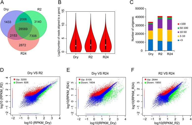
Fig. 4.

Summary of DGE-seq mapping data and comparison of expressed transcripts between dehydrated and rehydrated samples. a Numbers of shared and unique TACs among dehydrated and rehydrated gametophores. b Violin plot for the number of reads uniquely mapped to a TAC. c Number of transcripts with different expression levels in dehydrated and rehydrated gametophores as measured by DGE sequencing. d-f The scatter plot comparing the gene expression levels pairwise among the three libraries (between Dry and R2, between Dry and R24, as well as between R2 and R24, respectively). The number of DEGs were also present in the figure
