Figure 3.

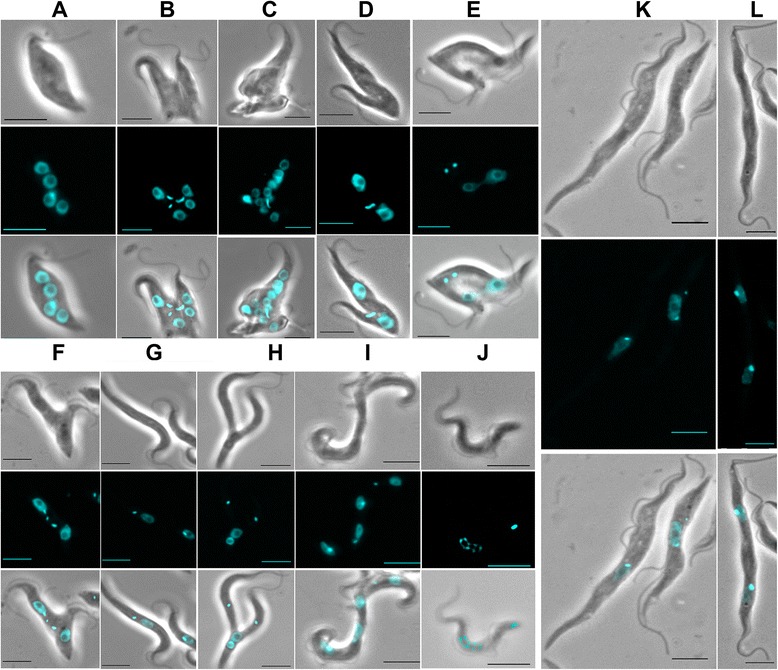
Pleiotropic morphological changes associated with growth defects following RNAi knockdown in T. brucei. A: Example of a multinucleated cell (observed in the mutant cell line T286). B: a multinucleated cells showing aborted cell division and over-replication of the kinetoplasts (T286). C: a multinucleated cell showing a complete disorganization of the cell morphology (T286). D: a cell with an abnormally segregating kinetoplast (T194). E: a dividing cell showing mis-positioning of the kinetoplasts between the dividing nuclei, with impaired cytokinesis (T290). F: a dividing cell showing mis-positioning of the kinetoplasts, together with over-replication of the kinetoplasts and impaired cytokinesis (T287). G: elongated cells observed in the mutant line T293. H: a dividing elongated procyclic form showing abnormal positions of the kinetoplasts (both anterior) and nuclei (both posterior) (T293); a ‘zoid’ might originate from this abnormal division process. I: large and elongated multinucleated cells (T317). J: fragmented (‘apoptotic-like’) nuclei (T221). K: elongated cells observed in the mutant lines T177. L: elongated dinucleated cell with impaired cytokinesis (absence of cleavage furrow) with daughter cells still joined at their posterior end (T178). Bar: 5 μm.
