
Fig. 1.
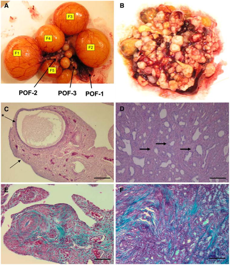
Anatomy and pathology of the hen ovary: (A) Gross anatomy of normal ovary showing follicular hierarchy (F1–F5); arrows point to postovulatory follicles (POF-1–3). (B) Ovarian tumor from hen with cancer confined to the ovary. (C) H&E stain of normal ovary, showing small developing follicle, (arrow points to OSE, *arrow points to granulosa cell layer). (D) H&E stain of ovary with cancer (arrows points to endometrioid-like gland), (E) Gomori trichrome stain of normal ovary; (F) Gomori trichrome stain of ovarian tumor. Calibration bar, 100 μm