
Fig. 2.
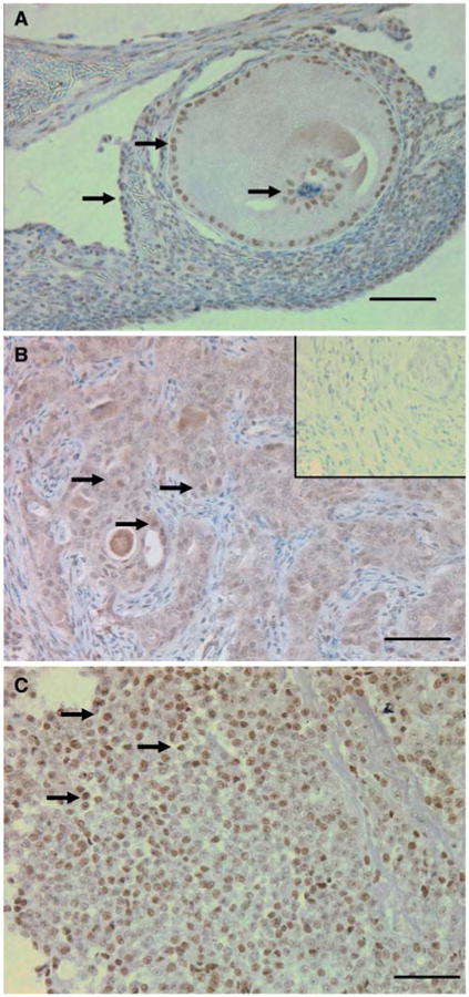
COX-1 immunohistochemistry: (A) Normal ovary (arrows point to COX-1 positive nuclei); (B) Ovarian tumor (arrows point to COX-1 positive nuclei), inset: non-immune IgG. (C) POF-1 from normal ovary, COX-1 expression is seen throughout the ovarian tumor whereas in the normal ovary COX-1 is confined to the granulosa cells and adjacent stroma. COX-1 is highly expressed in POF-1 (arrows point to COX-1 positive nuclei). Calibration bar, 50 μm