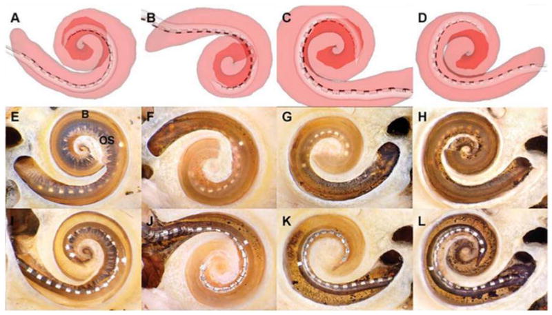
Figure 1.

Electrode arrays entirely within scala tympani (ST). In four temporal bones, the arrays remained within the ST. Representative screen captures for each of the reconstructions are illustrated in A–D. The ST is shaded translucent red, allowing the array to be seen inside (the scala vestibuli [SV] is not shown). The darker red area outlines the path of the apical turn. E–H are corresponding photographs of each cochlea after microdissection. In each case, the array can be seen through the basilar membrane (B) and osseous lamina (OS) to be resting entirely within the ST. In these specimens, the apical cochlear turn has been removed to provide an unobstructed view of the basal turn. I–L depict the same specimens after removal of the osseous lamina and basilar membrane, allowing direct visualization of the arrays in the ST.
