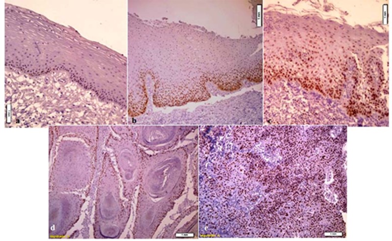
Figure 1.

a: MCM3 immunoreactivity in normal mucosa; MCM3 expression is restricted to nuclei of basal cell layer and a few cells in the immediate suprabasal layers. b: MCM3 immunoreactivity in mild epithelial dysplasia; MCM3 expression is restricted to the lower third of the epithelium. c: MCM3 immunoreactivity in severe epithelial dysplasia; MCM3 positively stained nuclei are present throughout the epithelium. d: MCM3 immunoreactivity in well-differentiated SCC; there are unstained foci of differentiating cells adjacent to keratin pearls. e: MCM3 immunoreactivity in poorly differentiated SCC; MCM3 expression is scattered throughout the malignant epithelial cells
