Figure 4.
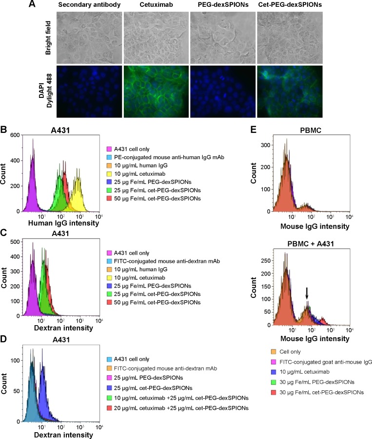
Binding specificity of cetuximab, PEG-dexSPIONs, and cet-PEG-dexSPIONs.
Notes: (A) For immunostaining of A431 cells, the cells were incubated with the indicated antibody or particles on ice for one hour. Dylight 488-conjugated anti-human Fcγ antibody was used as the detection antibody for fluorescence imaging. The nuclei of the cells were counterstained with DAPI (blue color). Bright field and merged cell images of Dylight 488/DAPI are shown in the upper and bottom rows, respectively. Images were obtained with a 40× objective lens. (B, C) Flow cytometry analysis of antibodies or nanoparticles specific to A431 cells. A431 cells were incubated with human IgG (negative control), cetuximab (positive control), or the synthesized magnetic probes at the indicated concentrations on ice for one hour. The cells were then stained on ice in the dark with R-PE-conjugated mouse anti-human IgG mAb (B) and fluorescein isothiocyanate-conjugated mouse anti-dextran mAb (C) for one hour to detect the human IgG molecules and dextran-coated magnetic nanoparticles, respectively, on the surface of A431 cells. (D) Competition assay by flow cytometry. The competition assay was performed as described in (C) except that A431 cells were treated on ice for one hour with 10 or 20 μg/mL cetuximab prior to treatment with the iron oxide nanoparticles. (E) Flow cytometry results showing that cet-PEG-dexSPIONs specifically targeted A431 that was spiked into PBMCs (105 A431 cells were spiked into 106 PBMCs). Fc receptors on the surface of PBMCs were blocked with FcR blocking reagents at 4°C for 20 minutes, and the cells were then incubated with cetuximab (positive control) or the synthesized magnetic probes at the indicated concentrations on ice for one hour. The cells were then immediately stained on ice in the dark with fluorescein isothiocyanate-conjugated goat anti-mouse IgG secondary antibody for an additional 30 minutes to detect cetuximab molecule binding to the surface of A431 cells. The arrow indicates the signal for unstained A431 cells.
Abbreviations: cet, cetuximab; DAPI, 4′,6-diamidino-2-phenylindole; dex, dextran; Ig, immunoglobulin; PEG, polyethylene glycol; SPIONs, superparamagnetic iron oxide nanoparticles; PBMCs, peripheral blood mononuclear cells; PE, phycoerythrin; mAb, monoclonal antibody.

