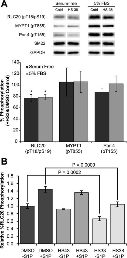
Figure 3.
Effects of HS38 and HS43 on phosphorylation in cells. (A) Human coronary artery smooth muscle cells (CA-VSMCs, passage 12) were serum-starved overnight, incubated for 40 min with HS38 (10 µM) or vehicle control (DMSO), and treated or not with 5% FBS for 2 min prior to lysis in SDS-PAGE buffer and Western blotting with anti-pT18/S19-RLC20, anti-pT855-MYPT1, or anti-T155-Par-4. Phosphorylated bands were quantified by scanning densitometry and normalized to the loading control (SM22). Phosphorylation levels in the presence of HS38 are expressed as a percentage of control (absence of ZIPK inhibitor). Values represent means ± SEM for n = 6 or 7 separate treatments. *Significantly different from control treatment (Student’s t test, p < 0.05). (B) Serum-starved aortic SM cells were treated with S1P (0.25 µM) and HS38 (±50 µM) or HS43 (±50 µM) for 30 min. HS38 significantly decreased the relative percent RLC20 phosphorylation at the basal (unstimulated) state (p < 0.0002, n = 8) and S1P stimulated state (p < 0.0009, n = 8), while HS43 did not significantly affect phosphorylation.

