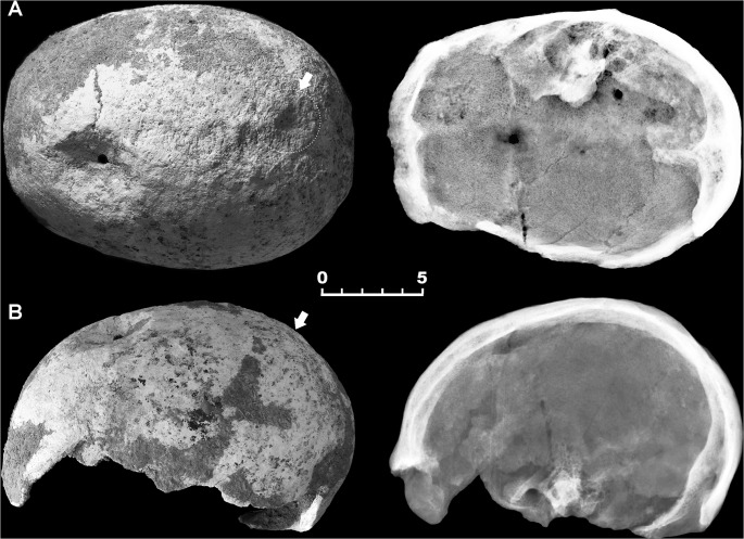
Fig 2. Double skull trephination affecting specimen 6589.1.
Superior (A) and lateral (B) view and corresponding X-ray images of the skull-cap. Wide crater-like elliptical depression across the bregma, showing well-defined, irregular borders (A, B). Gross morphology and X-ray images show healing, new bone being more radiolucent and with less mature architecture compared with surrounding old bone. A further minor depression in located close to the lambda (arrows) (A, B). Scale bar equals 5 cm.

