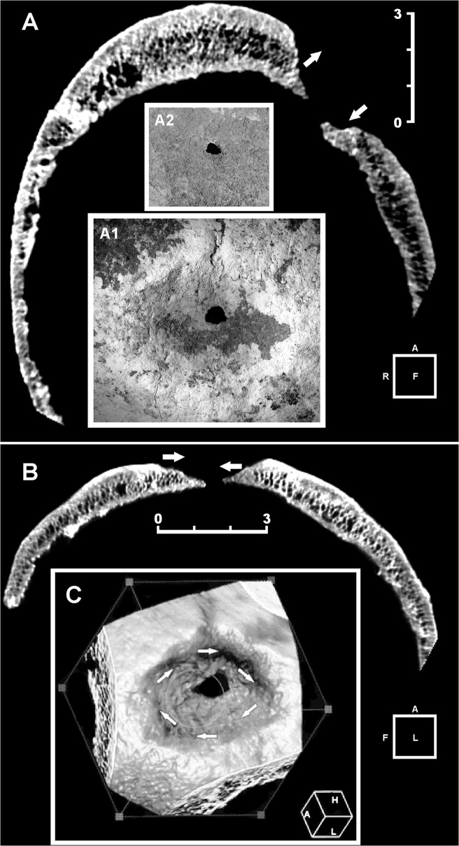
Fig 3. Three-dimensional (3D) computed tomography (CT) scan of the main skull trephination.
Frontal (A) and sagittal (B) plane CT scan. Extensive and irregular bone reconstruction is apparent. No evidence is seen of incomplete healing processes as in the case of complication by infection. Note the different angle at the top/bottom (frontal plane) (A) and front/back (sagittal plane) (B) bone around the hole, indicative of right-handed clockwise rotation applied during the action of scraping (arrows). Detail of the main lesion, whose edges show active regenerative bone processes (A1). Endocranial aspect of the lesion. The bone surrounding the hole is intact (A2). 3D reconstruction of the outer aspect of the trepanned cranial vault. The arrows show the clockwise rotation movement (C). Scale bars measure 3 cm.

