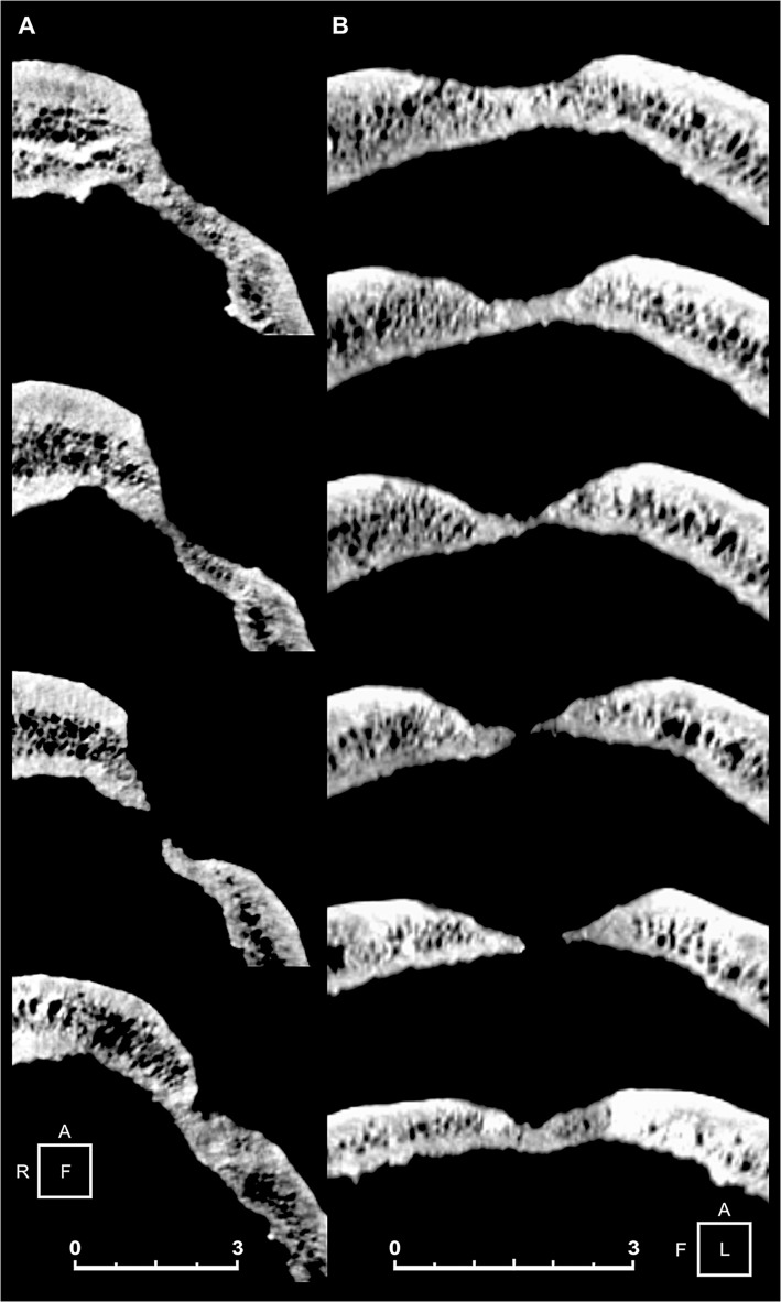
Fig 4. Three-dimensional (3D) computed tomography (CT) scan slices of the main skull trephination.
Coronal (A) and sagittal (B) plane CT scan cross section sequence. Note the oblique orientation of the hole walls, and the defect edges remodeled into one compact bone layer, as a result of the loss of diploic structures. The smoothed, beveled edges are also indicative of bone regrowth. Scale bars measure 3 cm.

