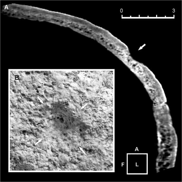
Fig 5. Three-dimensional (3D) computed tomography (CT) scan of the minor skull trephination.
(A, B) Sagittal plane CT scan (A). Only the external cortical layer of the bone is affected (arrow). Detail of the cone-shaped lesion, identified as an incomplete trephination with a possible ritual purpose (B). Bone remodeling, hypervascularity and pitting of the ectocranial surface suggest an advanced phase of the bone healing process. Scale bar measures 3 cm.

