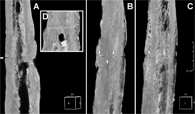
Fig 8. Three-dimensional (3D) computed tomography (CT) scan of the femoral lesion.
Posterior (A), anterior (B), and medial (C) views of the transverse simple linear fracture. New bone apposition (involucrum) is evident. The bone lesion is characterized by poor healing (arrows) (D). Detail of the cloaca opening (box) and, above, the uninterrupted fracture line. Scale bar equals 3 cm.

