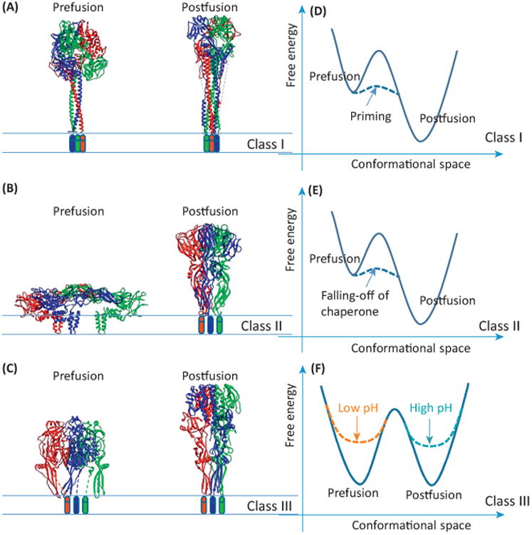
Figure 1.

The three classes of viral fusion proteins. (A–C) Side views of the viral fusion proteins in their prefusion and postfusion conformations for (A) class I, (B) class II, and (C) class III. The atomic models are shown as ribbons and the three monomers of each model are colored differently. Cylinders represent transmembrane helices whose structures are unknown. (D–F) Free energy landscapes of the three classes of viral fusion proteins for (D) class I, (E) class II, and (F) class III. Unbroken lines: landscapes of original proteins or assemblies; broken lines: landscapes of proteins after specific event as marked. Coordinates in these three panels are for illustrative purpose only and are arbitrary.
