Figure 3.
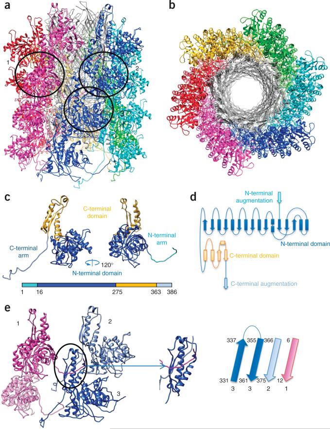
Structure of the pyocin sheath. (a,b) Side (a) and top (b) views of the atomic model of the pyocin trunk. Sheath subunits are shown in colors, and the tube is shown in gray. (c) Ribbon structure of the sheath monomer. Additional ribbon diagram is in Supplementary Video 6. (d) Topological diagram of the sheath monomer. (e) Joining of three (with numbers matching those in a) of the four adjacent monomers of the sheath protein, via β-sheet augmentation in their C domains (oval and inset). The polarities and identities of the β-strands in this augmented sheet are illustrated on the right.

