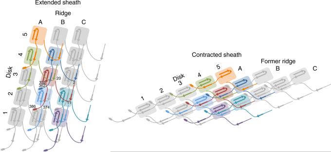
Figure 4.
Schematic diagram for the pyocin sheath topology of the extended mesh created by the N- and C-terminal extension arms within the sheath in the pre- and postcontraction states. β-strands participating in the sheet augmentation of the C domain are shown. α-helices involved in intersubunit interactions are shown as rectangles. Residue numbers for the subunit in red are given for strategic locations.

