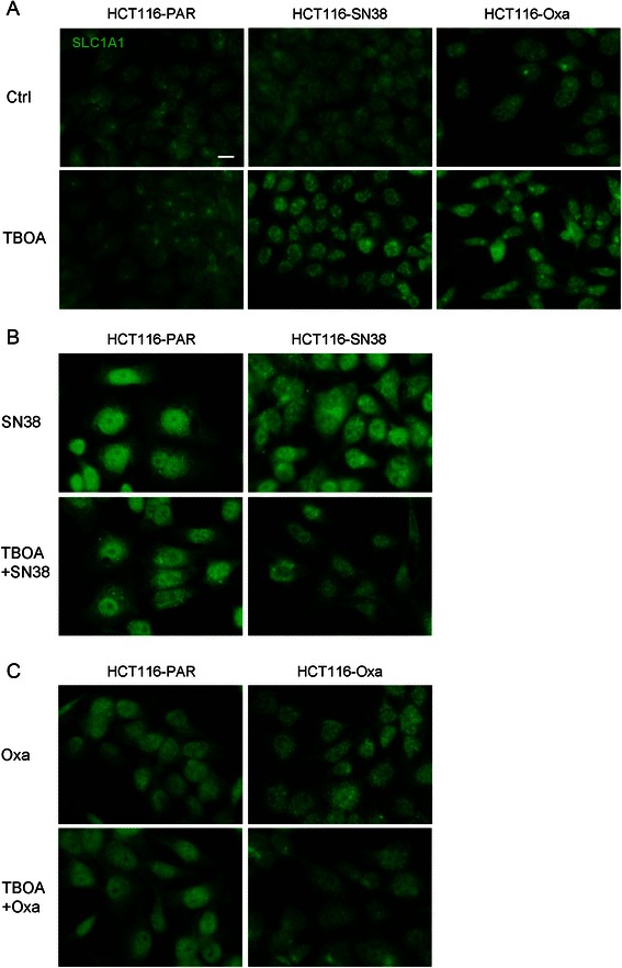
Fig. 7.

Subcellular localization of SLC1A1 in parental and resistant CRC cells – effects of chemotherapy and DL-TBOA. a Immunofluorescence images of parental (PAR), SN38 resistant and oxaliplatin resistant HCT116 cells treated or not for 48 h with 350 μM DL-TBOA, and stained with antibody against SLC1A1. b Parental and SN38-resistant HCT116 cells treated for 48 h with 0.8 μM SN38 in the absence or presence of 350 μM DL-TBOA, and stained as in A. c Parental and oxaliplatin-resistant HCT116 cells treated for 48 h with 20 μM oxaliplatin in the absence or presence of 350 μM DL-TBOA, and stained as in A. All conditions are representative of 2 or 3 independent biological replicates in duplicate. Scale bar: 10 μm. Additional file 7: Figure S7 shows the same images, merged with staining for DAPI and Rhodamine-conjugated phalloidin to visualize localization of nuclei and F-actin, respectively
