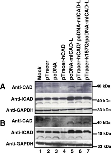
Fig. 3.

ICAD expression enhances CAD expression. Two sets of SUNE1 cells were transiently transfected with vector pTracer (Lane 2), pcDNA (Lane 3), pTracer-hCAD (Lane 4), pcDNA-mICAD-L (Lane 5), or cotransfection with pTracer-hCAD/pcDNA-mICAD-L (Lane 6) and pTracer-hCAD (K157Q)/pcDNA-mICAD-L (Lane 7). A mock transfection (No DNA) was included as control (Lane 1). Transfected cells were either not treated (Panel a) or treated with 50 μM of H2O2 for 6 h at 17 h post-transfection (Panel b). Protein was extracted as detailed in Materials and Methods. Expression of CAD, ICAD and GAPDH was each analysed by anti-CAD, anti-ICAD and anti-GAPDH respectively
