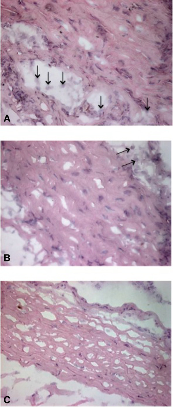
Figure 4.

Scleral inflammatory response and histological integrity. Scleral tissues (400×). (A) In group 1, moderate inflammatory response characterized by the presence of lymphocytes (↑) and loss of the arrangement of the collagen fibers are shown. (B) In group 2, few lymphocytes (↑) and a slight alteration in the histological integrity were observed. (C) In group 3, cells involved in the inflammatory response are not shown, and the arrangement of the collagen fibers was restored, which means that the scleral histology was recovered.
