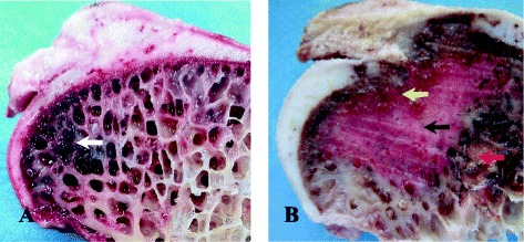
Fig. 5.

The comparing of normal and postoperative ostrich femur head. a The vertical plane of normal femoral head: the subchondral bone had no interruptions, and the bone marrow tissue was present within the bone trabecula (white arrow). b The vertical plane of collapsed femoral head postoperative: the subchondral bone was the yellow arrow, and the inflammatory pseudotumor the red arrow
