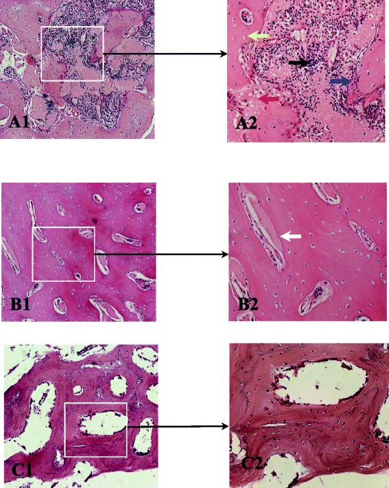
Fig. 6.

The result of H&E staining findings. a The H&E staining of the necrotic areas (A1 by 100 light microscope, A2 by 200 light microscope): the empty lacunae in the necrotic trabecular bone (yellow arrow). The irregular edges of the trabecular bone became due to bone resorption (blue arrow). The fibrous tissue within trabecular bone (black arrow) and fat cell (red arrow). b The sclerotic areas of trabecular bone (B1 by 100 light microscope, B2 by 200 light microscope): the trabecular bone thickened, the space of trabecular bone narrowed, and the nuclei of the active bone proliferation (white arrow). c The morphology of the normal trabecular bone: the uniform thickness and the space of trabecular bone and the normal morphology of bone marrow tissue
