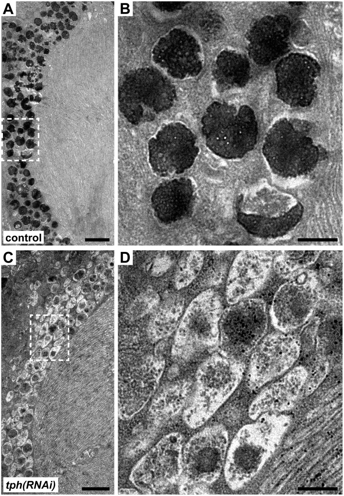
Fig 3. tph(RNAi) animals regenerate pigment cup cells largely devoid of mature melanosomes.
All micrographs are transverse sections of the planarian eye. (A) Electron micrograph of the photoreceptor rhabdomes and the pigment cup cells in control RNAi animals. Panel (B) is a magnified view of the pigment cup cells highlighting the mature melanosomes. (C-D) Electron micrograph of the photoreceptors of tph knockdown animals. In tph knockdowns the photoreceptors and the pigment cups are intact, but the melanosomes appear immature and less electron dense compared to controls. Scale bars in A and C are 2000 nm; in B and D are 500 nm.

