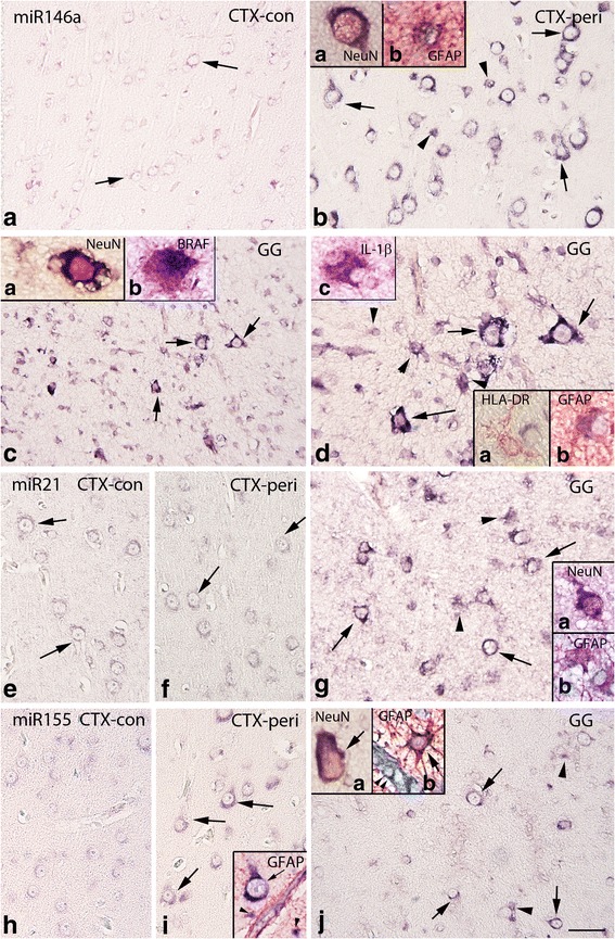
Fig. 3.

In situ hybridization of miR146a, miR21, and miR155 expression in control, peritumoral cortex, and GG. a–d miR146a. a Control cortex (−con); miR146a was expressed at low levels in neurons (arrows) and was undetectable in glial cells. b Peritumoral cortex (CTX-peri), showing miR146a expression in neurons (arrows) and glial cells (arrowheads). c–d GG; miR146a was expressed in both the neuronal (arrows) and the glial (arrowheads in d) tumor components. Inserts (a) in (b) and (c) show expression of miR146a in a neuron (NeuN positive, red); inserts (b) in (b) and in (d) show colocalization (purple) of miR146a with GFAP (red). Insert (b) in c shows colocalization (purple) of miR146a with BRAF (red). Insert (a) in (d) shows absence of colocalization with HLA-DR (microglia, red). Insert (c) in (d) shows colocalization (purple) of miR146a with IL-1β (red). e–g miR21. In both control (e) and peritumoral cortex (f), miR21 was expressed at low levels in neurons (arrows) and was undetectable in glial cells. g GG, showing miR21 expression in neurons (arrows) and glial cells (arrowheads). Insert (a) in (g) shows expression of miR21 in a neuron (NeuN positive, red); insert (b) shows expression of miR21 in astrocytes (GFAP positive, red). h–j miR155. h Control cortex, showing low expression of miR155. In peritumoral cortex (i), moderate expression was observed in neurons (arrows). j GG, showing miR155 expression in neurons (arrows) and glial cells (arrowheads). Insert in i shows expression of miR155 in a neuron (arrow) and astrocytes (GFAP positive, red; arrowheads) around a positive blood vessel. Insert (a) in j shows expression of miR155 in a neuron (NeuN positive, red); insert (b) shows expression of miR155 in an astrocyte (GFAP positive, red; arrow) around a positive blood vessel (arrowheads); scale bar in (j): (a, c) 150 μm; (b, e–j) 80 μm; (d) 40 μm
