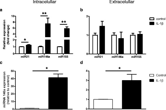
Fig. 5.

miRNAs expression levels after exposure to IL-1β in culture. Quantitative real-time PCR. Cellular (a) and extracellular (b) levels of miR21, miR146a, and miR155 in U373 glioblastoma cells 24 h after exposure to IL-1β (10 ng/ml). Cellular (c) and extracellular (d) levels of miR146a in human fetal astrocytes in culture 24 h after exposure to IL-1β (10 ng/ml). Data are expressed relative to the levels observed in unstimulated cells and are mean ± SEM from two cultures (*p < 0.05 compared to control). miRNAs expression was normalized to that of the U6B small nuclear RNA gene (Rnu6B) or to that of miR23a for the intra- and extracellular fractions, respectively
