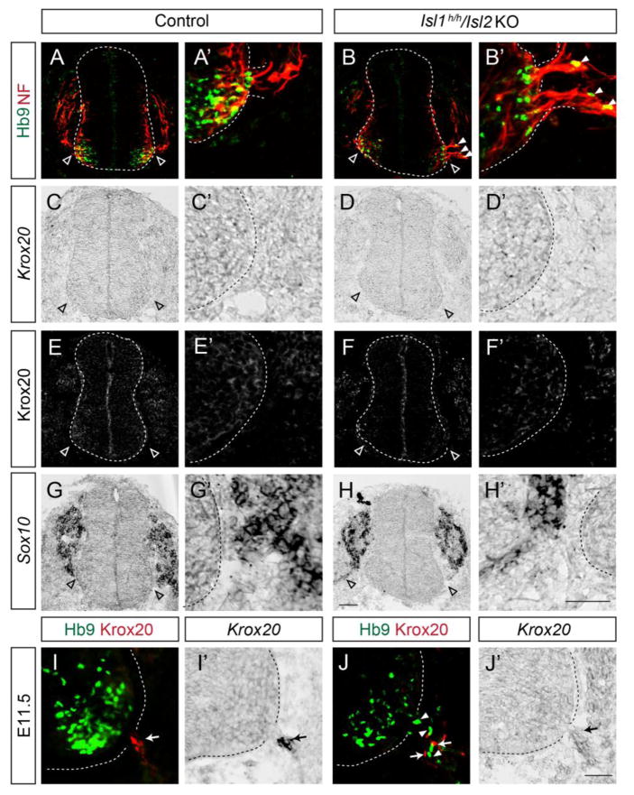
Figure 3. Motor somata exit the neural tube before the settlement of boundary cap (BC) cells in Islet mutant spinal cords.
(A–B′) Motor neurons (Hb9, green) are ectopically located in the ventral roots (Neurofilament, red) of Isl1 hypo; Isl2 KO mice at E9.75. (C–H′) BC cells labeled by Krox20 protein or mRNA as well as migrating neural crest cells labeled by Sox10 mRNA are absent in the dorsal and ventral exit points in adjacent sections of A and B. Empty arrowheads indicate MEPs (A–H′). (I–J′) At E11.5, BC cell clusters are present at MEPs in control mice (arrows, I, I′), while scattered BC cells (arrows, J, J′) intermingled with ectopic motor neurons (arrowheads, J) are seen in Isl1 hypo; Isl2 KO mice. Expression of Krox20 was assessed by immunohistochemistry and in situ hybridization in adjacent sections. Scale bars: 50 μm.

