
Figure 7.
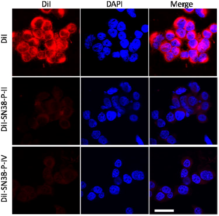
Confocal fluorescence microscopy images of HT-29 cells incubated with 1 μg/mL of free DiI and DiI-SN38 co-loaded micelles at 37 °C for 2 h. Cell nuclear was stained with DAPI (Blue). Scale bar: 30 μm.
Official websites use .gov
A
.gov website belongs to an official
government organization in the United States.
Secure .gov websites use HTTPS
A lock (
) or https:// means you've safely
connected to the .gov website. Share sensitive
information only on official, secure websites.
Confocal fluorescence microscopy images of HT-29 cells incubated with 1 μg/mL of free DiI and DiI-SN38 co-loaded micelles at 37 °C for 2 h. Cell nuclear was stained with DAPI (Blue). Scale bar: 30 μm.