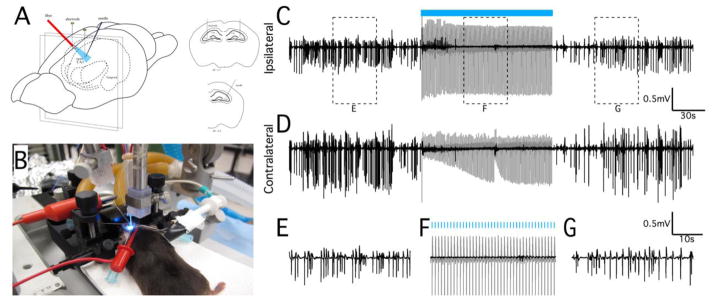
Figure 1.

Optical stimulation at 1Hz can suppress in vivo epileptiform activity. (A) Schematic representation of in vivo optical stimulation in the mouse hippocampus. Actual experimental preparation is shown in (B). Recording electrodes were positioned bilaterally in the septal CA3 regions. Epileptiform activity was induced by unilateral bolus injection of 40mM 4-AP, and an optical fiber was positioned to maximally illuminate the dorsal hippocampus near the injection site. Neural recordings from electrodes positioned in the ipsilateral (C) and contralateral (D) septal CA3 regions showing hippocampal seizure activity before, during, and after a 2 min period of optical stimulation at 1Hz (indicated by blue bar). Stimulation was only applied to the site of 4-AP injection (ipsilateral site), but reduction of epileptiform activity occurred bilaterally. (E–G) Magnification of the regions identified in (C) to show the seizure and suppressed activity in greater detail. Note that the optical evoked potential during the stimulation period (gray traces) was removed from these recordings by template subtraction (black trace, see Methods). Stimulation trials in wild-type control animals did not elicit an optical evoked potential and had no effect on epileptiform activity (data not shown).
