Figure 10.
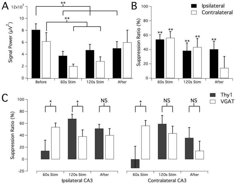
LFS optical suppression of epileptiform activity in vivo by driving solely hippocampal interneurons. (A) Signal power calculated before, during, and after 2-minute 1Hz optical stimulation of adult VGAT mice during ongoing electrographic seizure. Data is shown for both the ipsilateral and contralateral recording sites. Statistically significant reduction in signal power as compared to baseline was observed ipsilaterally in all measurement periods as well as contralaterally during stimulation. (B) Suppression ratio comparison of measurement periods, showing a 40%–60% reduction in seizure activity as a result of LFS of ChR2-expressing interneurons. (C) Summary of responses to oLFS to compare suppression efficacy in the Thy1- and VGAT-driven transgenic mouse lines. While stimulation in VGAT animals produced more seizure reduction than in Thy1 animals at the beginning of stimulus trials, the amount of seizure suppression that could be achieved with prolonged stimulation was greater in Thy1 animals. Results are mean ± SEM (n=7 Thy1, n=9 VGAT). *P≤0.05, **P<0.01, Student’s t-test.

