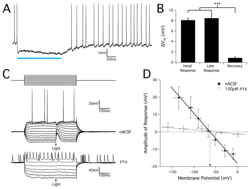
Figure 8.
Optical response in juvenile VGAT hippocampal slices is GABAA-mediated and causes pyramidal cell hyperpolarization in normal conditions. (A) Hyperpolarization response in pyramidal cells stimulated with a 1s sustained optical pulse. Spontaneous cell firing was completely inhibited during the optical stimulus. (B) Measurement of the magnitude of optically-induced change in membrane potential from hippocampal interneuron activation. Though membrane voltage often continued to hyperpolarize slightly, the amount of cell hyperpolarization did not change significantly over the course of the stimulus pulse. After optical stimulation, cells fully recovered to their baseline resting membrane potential. Results are mean ± SEM, n=27. P<0.001, one-way ANOVA. (C) Example pyramidal cell response to a 10ms optical pulse stimulus during a series of hyperpolarizing and depolarizing injected current pulses (top traces). Optical stimulus is marked by an arrow. The stimulus protocol was repeated after applying the GABAA-blocker PTX (100μM), which completely eliminated any optical response. (D) Amplitude of optical response was measured and plotted against the membrane potential for each current pulse in order to determine the reversal potential for the optical response. After linear regression, the potential at a zero-response was calculated to be at −63.3±0.70 mV (mean ± SEM, n=23, data point error bars represent SD). In 100μM PTX conditions, response amplitudes were nearly zero for all membrane voltages with no clear reversal potential that could be identified.

