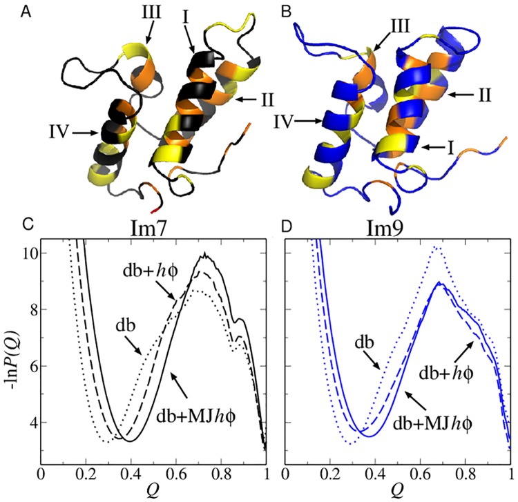
Fig 1. Structures and folding thermodynamics of Im7 and Im9.
The ribbon diagrams (top) depict the PDB structures of (A) Im7 (PDB ID: 1AYI) and (B) Im9 (PDB ID: 1IMQ). The positions of four types of strongly hydrophobic residues (M, F, I, L) are shown in orange whereas those of four types of largely nonpolar residues but have weaker hydrophobicities (V, W, Y, A) are shown in yellow. Other residue positions are shown in black (for Im7) or blue (for Im9). Each structure contains four helices (I, II, III, and IV). The bottom panels show free energy profiles −ln P(Q) for Im7 (C) and Im9 (D) computed using three different models around each model’s transition midpoint.

