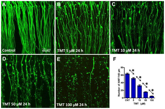
Figure 2.
Representative confocal images showing the condition of the peripheral auditory nerve fibers (ANF) from the upper middle turn of the cochlea. ANF in control group (A) and groups treated with indicated dose of TMT 24 h (B–E). (F) Mean (+SEM) numbers (n = 14/group) of ANF/140 μm from the upper middle turn of the cochlea in control cultures and cultures treated with various concentrations of TMT for 24 h. Individual groups significantly different from one another (* p < 0.05).

