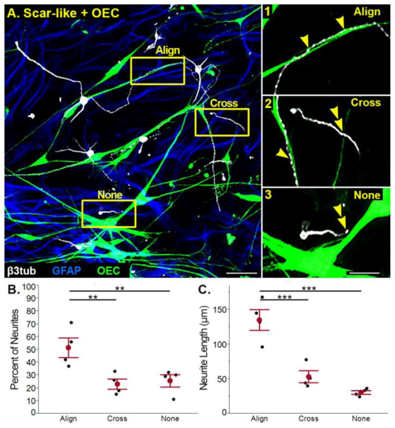
Figure 4. OEC-enhanced neurite outgrowth is mediated by neuron-OEC association.

(A) In scar-like + OEC cultures associations between neurons and OECs were classified as: 1) aligned (single arrowheads), 2) crossing (double arrowhead), or 3) no interaction (double arrowhead). (B) An average of 52% of measured neurites aligned with OECs, while 23% of neurites crossed and 26% did not associate with OECs. (C) Neurites aligned with OECs were longer than those that crossed or did not associate with OECs. Individual experiments (n=4) in B and C are represented by black dots, with the means ±SEM values marked in red. GFAP, Glial fibrillary acidic protein; β3-tub, β3-tubulin; OEC, olfactory ensheathing cell. Scale A = 50 μm; A1–3 = 25 μm.
