
Fig. 5.
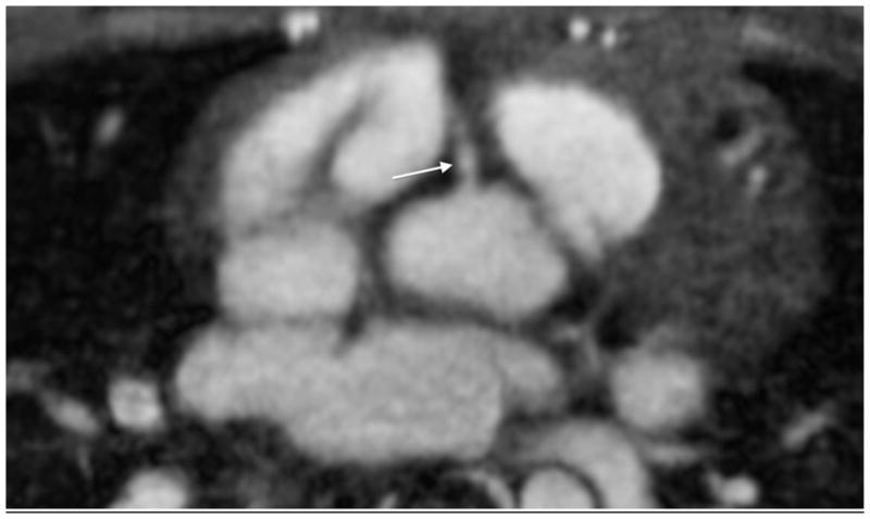
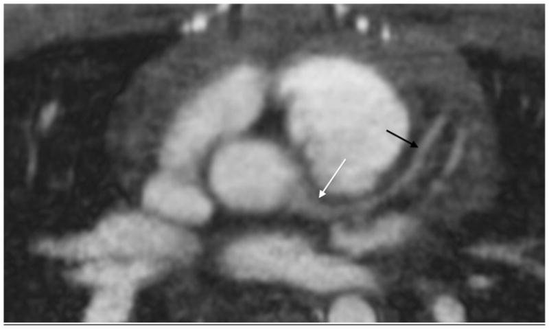
A 10-year-old boy with pulmonary artery stenosis. a Gated axial spoiled gradient echo image at the level of the right coronary artery (arrow) shows its origin. b Axial image at level of the left main coronary artery (white arrow) also shows left anterior descending coronary artery (black arrows). The coronary vessels are not typically seen with spoiled gradient echo sequences with such clarity