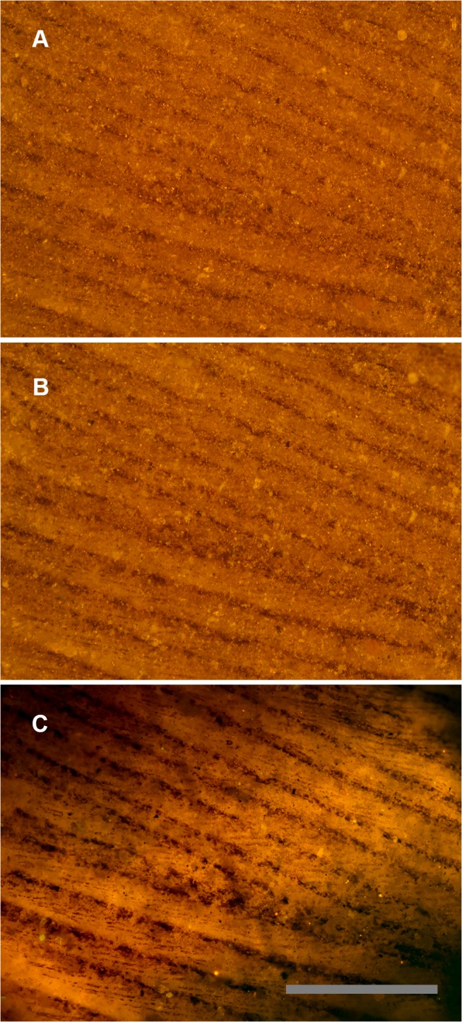
Fig 3. Feather under reflected and matrix fluoresced illumination.

Green River Formation feather using identical images under different lighting conditions. A, Reflected light microscopy, only barbs are visible. B, Polarized light, some traces of barbules. C, Laser-stimulated fluorescence of matrix behind the carbon film backlights the feather and renders barbules visible across the entire field of view. Scale bar 0.5 mm.
