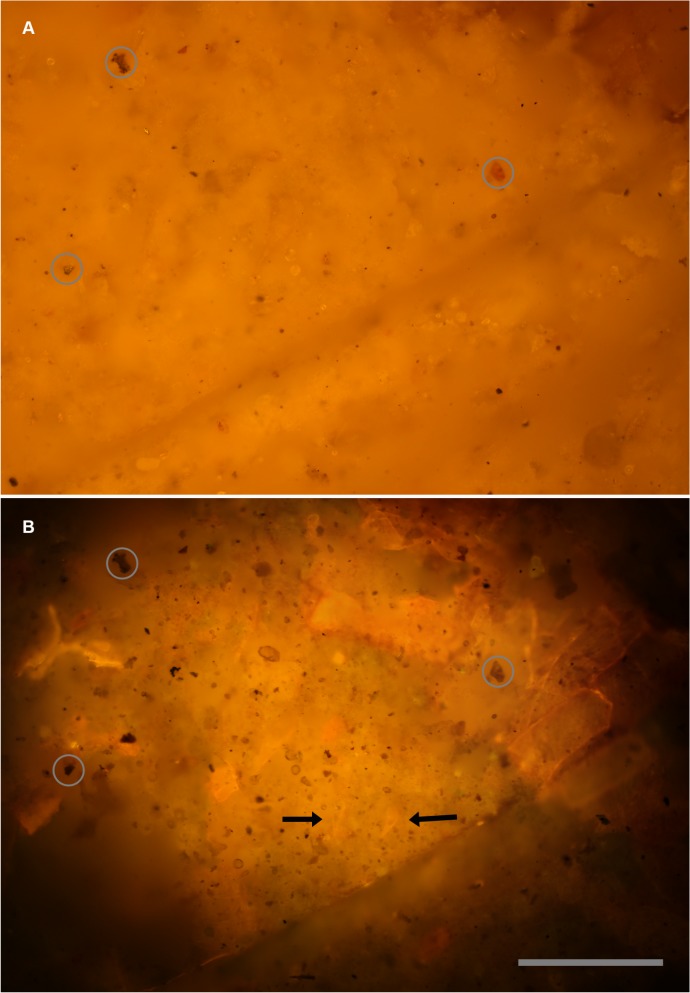
Fig 5. Micro-imaging—visible light vs.
laser fluorescence. Identical views of the same field. A, Reflected white light image of a purposely non-descript field. B, Same field illuminated with a 532 nm laser. Some features are at the specimen’s surface, but lack color contrast, others are visible within the matrix of the specimen. Black arrows indicate teeth found coincidentally in this image. Circles indicate reference landmarks. Scale bar 0.15 mm.

