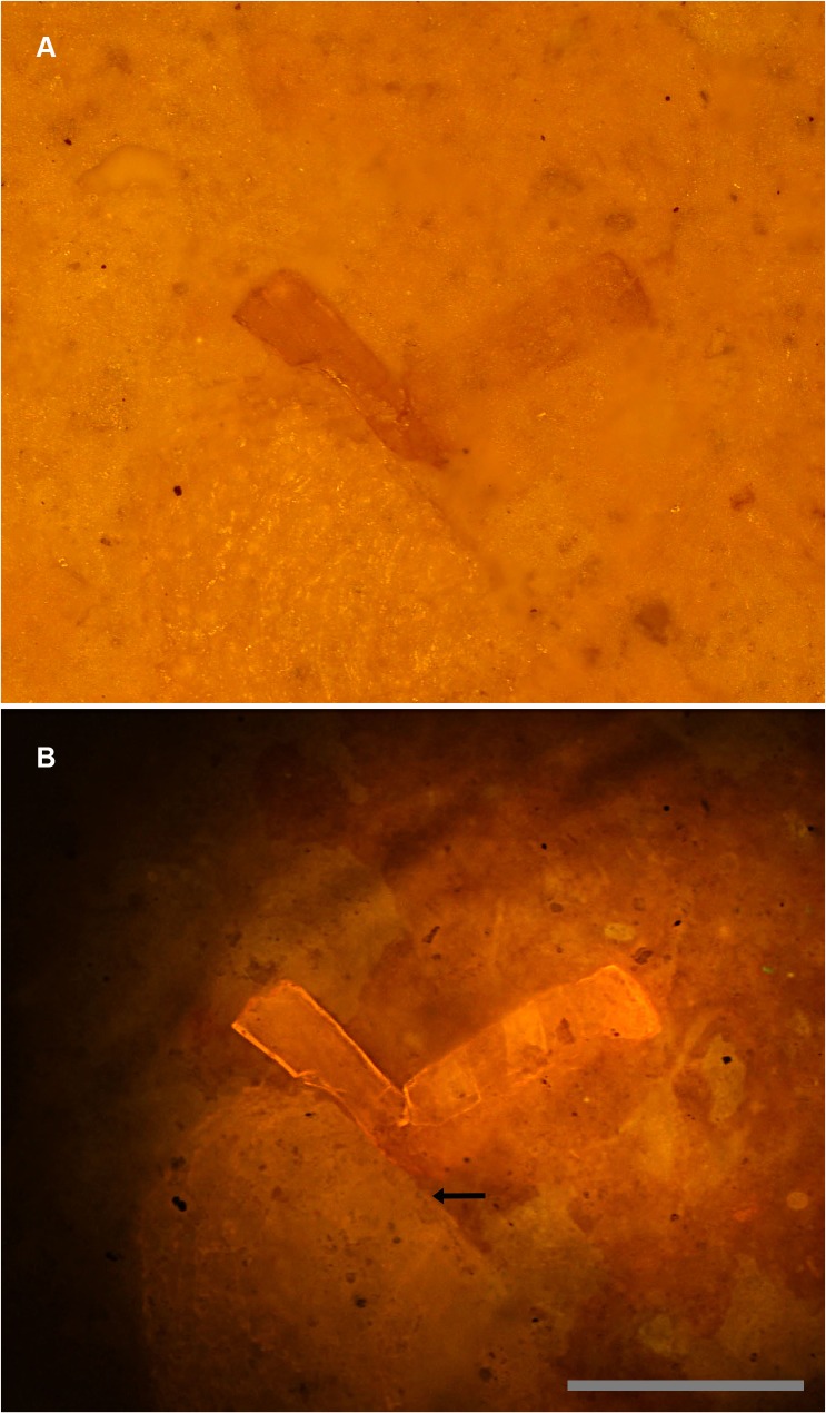
Fig 8. Sub-surface imaging.
A, White light micrograph shows the bone fragment on the right entombed within matrix. B, Specimen under laser fluorescence. Photograph shows a high level of detail invisible under white light. Note that another much larger fragment also becomes visible (arrow). Scale bar 0.5 mm.

