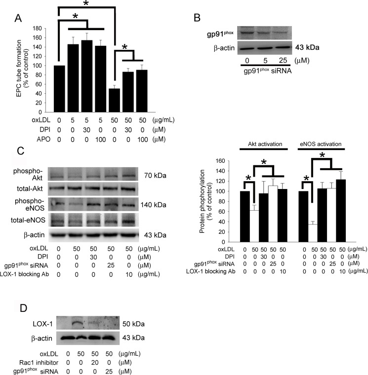
Fig 5. The NADPH oxidase-related pathway contribute to decreased tube formation in EPCs under high concentrated oxLDL.
(A) EPCs were pretreated with DPI or APO for 1 hour prior to treatment with 5 or 50 μg/mL oxLDL for 24 hours. In vitro angiogenesis was assayed using ECMatrix gel. Data were expressed as the mean ± SEM of three experiments performed in triplicate. *p < 0.05 was considered significant. (B) EPCs were transfected with gp91phox siRNA, the total gp91phox protein were analyzed using western blotting. (C) EPCs were pretreated with DPI, LOX-1 blocking antibody for 1 hour or transfected with gp91phox siRNA prior to 50 μg/mL of oxLDL treatment, subsequently eNOS and Akt activation (phosphorylation) were analyzed by Western blot. Total eNOS, Akt, and β-actin protein levels were used as loading controls. The graph showed the quantitative activation of eNOS (phospho-eNOS/total-eNOS ratio) and Akt (phospho-Akt/total-Akt ratio) density in oxLDL-treated EPCs. (D) EPCs were pretreated with 20 μM Rac1 inhibitor for 1 hour or transfected with gp91phox siRNA prior to 50 μg/mL oxLDL treatment for 12 hours, subsequently membrane LOX-1 expression was analyzed by Western blot. β-actin protein levels were used as a loading control.

