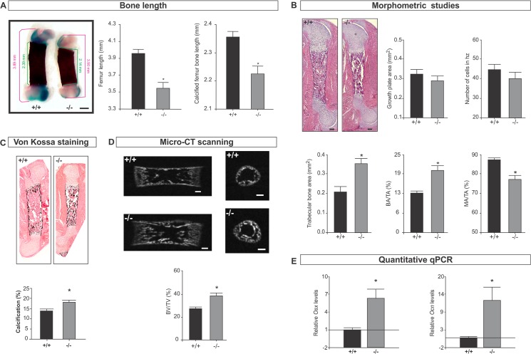
Fig 1. Femur bone development is affected in Spag17-deficient mice.
(A) Alcian Blue/ Alizarin Red staining of femur from wild-type (+/+) and Spag17-mutant (-/-) mice. Measurement of femur length and calcified femur bone length shows Spag17-mutant limbs are shorter than wild-type mice. Scale bars, 0.5 mm. (B) Histological and morphometric studies on femur shows increase bone formation in mutant mice. Scale bars, 200 μm. (C) von Kossa staining reveals increased calcification in mutant mice. (D) Micro-CT structural analysis on femurs; mid-sagittal sections (left) and cross-sectional trans-axial views at the mid-diaphysis (right). Images were acquired in 600 projections in 180 degrees of rotation with 500 miliseconds exposure per projection. Scale bars, 200 μm. (E) mRNA expression of osterix (Osx) and osteocalcin (Ocn) measured by qPCR. (+/+), wild-type; (-/-), Spag17-mutant mice. Data are presented as means ± SEM from ≥7 mice. * Indicates statistically significant differences, p< 0.05. hz: hypertrophic zone; BA/TA: bone area/total area; MA/TA: marrow area/total area; BV/TV: bone volume/total volume; Osx: osterix; Ocn: osteocalcin.

